Wednesday, November 24, 2010

Assignment 4

Paper: Bernard G. Steinetz, Laura T. Goldsmith, George Lust. (1987). "Plasma relaxin in pregnant and lactating dogs" Biology of Reproduction 37; 719-725.


Relaxin is a hormone secreted by the corpus luteum and placenta during the later parts of pregnancy. Relaxin has been shown in previous studies to allow more flexability in the pubic ligaments, vagina and cervix by decreasing collagen by stimulating collagenase enzymes. This study was designed to look at the detectable blood concentrations of relaxin, estradiol and progesterone during pregnancy and postpartum in two species of dogs.


Summary of the Article:

Researchers in this study followed weekly relaxin levels in the blood throughout
pregnancy, partuition and lactation in labrador retreivers and beagles. When females were in heat, they were placed with a male and allowed to mate. After copulation, weekly samples of relaxin, progesterone and estradiol concentrations were determined by double antibody radioimmunoassay. Blood
samples from male and female dogs that were not in heat, as well as females that had mated but not conceived (pseudopregnant) were used as controls.


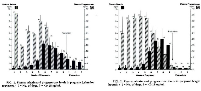
The results in figure 1 and 2 show that in the labrador retreiver relaxin levels begin to increase at 4 weeks
of pregnancy and peak at 6, then slowely decline. After 3 weeks postpartum however, levels are still higher than they were initially. This is in contrast to the levels in the beagle, which peak between 7-8 weeks, but return to initial levels in the following 3 weeks postpartum.


Figure 4 shows the levels of progesterone and estradiol during the study. They show that their patterns of secretion into the blood do not match that of relaxin. Progesterone declined before relaxin did, and estradiol levels were very low toward the end of the pregnancy while relaxin was still higher. Meanwhile Figure 3 shows the following 10 weeks postpartum, during which lactation occurs. The levels of relaxin in the beagle remain low and steadily decrease. The levels of relaxin in the labrador retreiver are much higher initially, but steadily decrease in the remaining weeks.


Finally, figure 5 shows progesterone and relaxin concentration in pseudopregnant dogs.The levels of relaxin in the blood remain undetectable during the 11 weeks they were measured. This shows that relaxin has a role in pregnancy, and is not secreted in response to psychological stimulus, or the act of copulation.


Critique:
  • This paper had good evidence suggesting the increase in levels of relaxin toward the middle and end of pregnancy in dogs. It also had good evidence to suggest that estradiol and progesterone and secreted independantly of relaxin. The figure showing pseudopregnancy was effective in showing that this hormone is secreted in response to changes that occur in pregnancy.
  • However, in this study, the sample size was very small, as it included only 5 beagles and 8 labrador retreivers, 3 of which did not become pregnant.


  • The blood concentrations of relaxin in the labrador retreiver were high after partuition and during lactation, while those of the beagle were not.The biggest problem with this experiment, and perhaps that which distorts alot of the data is the fact that 80% of the labrador retreivers used had hip dysplasia, a condition which possibly involves improperly functioning relaxin. If you are trying to determine normal levels of relaxin during pregnancy, it is not reliable to use a group of animals that possibly have a problem in regulating the hormone in question. Meanwhile, the beagle group which they compare the retrievers to had almost no incidence of hip displasia.


  • The authors say the fact that the labrador retreiver has higher base levels of relaxin is possibly related to the hip dysplasia, however to make such a statement, a control group without hip dysplasia would have to have been used. Furthermore, the relaxin antiserum R6, which they used for RIA is well documented in its effect of cross reacting with other relaxin-like peptides. This casts doubts on the identity of the hormone they are measuring after partuition in the labrador retreiver and possibly the entire experiment
Future experiments

The developmental stages of pregnancy in dogs needs to be investigated. In humans, the corpus luteum increases in size by 3x during the sixth week of pregnancy. If this is the case in the dog, this would coincide with the fact that relaxin is secreted by the corpus luteum.


More reliable methods of determining relaxin concentration after lactation are also needed, with a larger sample size.

Wednesday, November 10, 2010

The Function of Relaxin

Relaxin has been described by some researchers as a "fickle and elusive hormone" (Steinetz, 2000). While much has been discovered about relaxin, there is still much work to be done. Recent research, such as the collaborative work presented at the Relaxin conference (Geoffery W et al, 2000) show that relaxin is not only a pregnancy hormone, but has a wider range of collagen-related functions in the body.


Relaxin has been implicated in connective tissue remodeling, specifically by inhibiting collagen synthesis, while promoting matrix metalloproteinases, which degrade collagen. (Samuel S et al, 1998)


One of the best ways to determine the overall function of a hormone is to knock out its expression using transgenic animals. Transgenic rats have been used to study the effects of relaxin in areas such as the heart. High affinity relaxin receptors have been found in the aorta and heart chambers of both male and female rats (Osheroff L, Ho W., 1993). One study showed that knock out mice had a 30% increase in atrial weight, and moderate increases in the weight of other organs such as the lungs and liver (Samuel S et al, 2003). Increase in atrial weight caused by collagen build up resulted in impeded blood flood into the chambers, and reduced elasticity. While the rats did not show increased blood pressure, or heart rate, it is clear that relaxin acts to remodel collagen in body tissues.


During pregnancy, Relaxin and relaxin receptors have been found in areas such as the nipple, cervix, vagina and pubic symphysis. Knockout mice have been used to show that relaxin causes an increase in the length of pubic symphysis during the second half of pregnancy. However, it was shown that relaxin deficient mice can still deliver newborns, and the duration of gestation is unaffected. Similar results exist for the cervix and vagina of female rats. These studies have shown that relaxin increases the overall length or width, but is not necessary for a successful delivery (Ling Zhao et al, 1999). Interestingly, milk secretion seems to be dependant on collagen remodelling by relaxin. Knockout mice that delivered newborn babies were unable to feed their newborns with milk. While they produced milk, development of the mammary gland was not sufficient to secrete it (Zhao et al, 2000).


From the perspective of male physiology, relaxin has been shown to affect development of the male reproductive tract. Knock out mice showed increased weight in prostate glands, testis, epididymus and seminal vesicles. This lead to decreased growth and fertility, however the males still produced viable sperm. (Samuel CS,2003)


While the effects of relaxin are present in several areas of the body, the exact purpose of the hormone has been difficult to determine, as it has effects on collagen production in many tissues, but is not normally essential to these tissues. Despite this, it is produced in a wide variety of tissues including the heart, brain, corpus luteum, endometrium and ovaries. While the role of relaxin in many body tissues remains ambiguous, the development in the mammary gland does in fact seem dependant on relaxin.


REFERENCES


Samuel C.S., Coghlan J.P., Bateman J.F. (1998) Effects of relaxin, pregnancy and parturition on collagen metabolism in the rat pubic symphysis. J Endocrinol 159:117–125


Osheroff P.L., Ho W.H. (1993) Expression of relaxin mRNA and relaxin receptors in postnatal and adult rat brains and hearts. Localization and developmental patterns. J Biol Chem 268:15193–15199.


Xiao-Jun Du, Chrishan S Samuel, Xiao-Ming Gao, Ling Zhao, Laura J Parry, Geoffry W Tregear (2003) “ Increased myocardial collagen and ventricular diastolic dysfunction in relaxin deficient mice: a gender-specific phenotype” Cardiovasc Res (2003) 57 (2): 395-404.


Ling Zhao, Peter J. Roche, Jenny M. Gunnersen, Vicki E. Hammond, Geoffrey W. Tregear, E. Marelyn Wintour and Felix Becks (2000) "Mice without a Functional Relaxin Gene Are Unable to Deliver Milk to Their Pups Endocrinology" Vol. 140, No. 1 445-453


Zhao L., Samuel C.S., Tregear G.W., Beck F., Wintour E.M. (2000) Collagen studies in late pregnant relaxin null mice. Biol Reprod 63:697–703


Geoffery W Tregear, Richard Ivell, Ross A Bathgate, John D Wade “Relaxin 2000: The proceedings of the third international conference on relaxin & related peptides” Kluwer Academic Publishers


Samuel CS, Tian H, Zhao L, Amento EP, (2003) "Relaxin is a key mediator of prostate growth and male reproductive tract developement" Lab Invest, Jul;83(7):1055-67.

Tuesday, October 26, 2010

The Structure of my Least favorite Hormone: Relaxin


Relaxin is a 7 peptide hormone that is part of the insulin/IGF family. Similar to insulin, the active form of relaxin consists of two chains, linked by disulfide bridges. Studies show that no particular amino acid on the NH terminal region of the A chain is functionally important, but it is the presence of a helix that is required for biological activity (Erika et al.,1987).

RXFP1, RXFP2, RXFP3 and RXFP4 are the four receptors associated with Relaxin in humans (because I am finding it very hard to find information on this hormone with respect to other mammals). These are transmembrane G Protein coupled receptors. These glycosylated heptahelical domains are somewhat similar to FSH and LH receptors (shpakova, 2009).


Figure 1: Structural representation of Relaxin

Obtained from http://upload.wikimedia.org/wikipedia/commons/c/ce/Relaxin.png

To compare the similarity of Relaxin between species of Equus caballus (horse), Sus Scrofa (wild boar) and Felis catus (domestic cat), a sequence alignment was performed in ClustalW2, results are shown in the figure below.


Equus           -------------------------------IKACGRELARLRIEICGSLSWKKTVLRLE 29
Sus             MPRLFS-YLLGVWLLLSQLPREIPGQSTNDFIKACGRELVRLWVEICGSVSWGRTALSLE 59
Felis           MLRLFLSHLLGVWLLLSLRARKIP--AQEEVLKACGREFVRLQIRICGSLSWGKSSQQHR 58
                                               :******:.** :.****:** ::    .


Equus           EPGLEVGQPVEIVSSSISKDAEALNTKLGLNSNLPKEQKATLSERQPSWRELLQQPALKD 89
Sus             EPQLETGPPAETMPSSITKDAEILKMMLEFVPNLPQELKATLSERQPSLREL-QQSASKD 118
Felis           EPRQAPAALPEIVSSSITSGAEALNGMLEYIPDLPQELKATLSEREPSFREL--QPSLKD 116
                **    .   * :.***:..** *:  *   .:**:* *******:** ***  *.: **


Equus           SNLNLEEFEETIQKTQSEVEDDSLSELKNLGLDKHSRKKRMI--QLSHKCCYWGCT---- 143
Sus             SNLNFEEFKKIILNRQNEAEDKSLLELKNLGLDKHSRKKRLFRMTLSEKCCQVGCIRKDI 178
Felis           SNLNLEEVEKSILGRQNEAEDQSLSQLGRSRLDAHSRIKRSDYIRYSDRCCNVGCTRKEL 176
                ****:**.:: *   *.*.**.** :* .  ** *** **      *.:**  **    


Equus           ----
Sus             ARLC 182
Felis           ADLC 180

- * indicates identical residues
- :  indicates there are conserveral substitutions
- .  indicates substitutions are semi-conserved


The percent similarity between each species was compared, showing that the sequence is fairly conserved between species, between 56 and 66% similarity.

Table 1. Sequence similary of the hormone Relaxin between Equus caballus, Sus Scrofa (wild boar) and Felis Catus obtained in ClustalW2

SeqA Name    Len(aa)  SeqB Name    Len(aa)  Score
=================================================
1    Equus   143      2    Sus     182      66   
1    Equus   143      3    Felis   180      58   
2    Sus     182      3    Felis   180      56   
=================================================

References

Erika,E., Bullesbach, Christian,S. (1987) "Relaxin structure, quasi allosteric effect of the nhz-terminal a-chain helix" The American Society for Biochemistry and Molecular biology, 262(26)

Shpakov, Shpakova. (2009) "Low-molecular regulators of polypeptide hormone receptors containing LGR-repeats" Biomedical Chemistry 3(4); 351-360

Monday, October 11, 2010

Hormone of choice: Relaxin

Relaxin is a peptide hormone that is very similar to insulin in structure. This hormone plays an important role during pregnancy.
Female mammals give birth through the pelvis, a strong skeletal support structure of the lower limbs. At the bottom of the pelvis, two bones are joined together at the pubic symphysis. This is a cartilaginous joint that is relatively immovable. During birth, relaxin acts on this cartilaginous joint to allow the widening of the birth canal.


Figure 1. The Pelvic Girdle, with pubic symphsis labelled
http://ajs.sagepub.com/content/29/4/521/F10.large.jpg

Relaxin is produced in granulocytes in the corpus lutueum. It has also been observed in several other places including the placenta, uterus and endometrium. Relaxin has also been found in the testes of some male animals. In males, the role of this hormone is not known. In females, the overall effect is a more flexible, wider birth canal.
Research suggests that relaxin works by cutting collagen and inducing its breakdown in the pubic symphysis. A synthetic form has been developed for the treatment of scleroderma (a connective tissue disease that causes the skin to become tight and thick).


References
1. Anderson LL, Bast JD, Melampy RM. Relaxin in ovarian tissue during different reproductive stages in the rat. J Endocrinol 59:371–372, 1973

2. Sherwood OD, Crnekovic VE, Gordon WL, Rutherford JE. Radio-immunoassay of relaxin throughout pregnancy and during parturition in the rat. Endocrinology 107:691–698, 1980.

3. Perl E, Catchpole HR, (1950). "Changes induced in the connective tissue of the pubic symphysis of the guinea pig with estrogen and relaxin", Arch Pathol (chic), 50(2): 233-239